Figure 1.
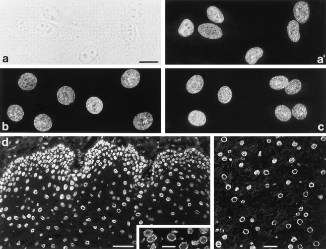
Immunofluorescence microscopy of cultured mammalian cells and tissue cryostat sections after reaction with mAb 203-37. (a) Phase contrast and (a′) epifluorescence optics of human adenocarcinoma cells of line HeLa. (b–e) Epifluorescence optics of cultured bovine mammary gland epithelial cells of line BMGE (b), porcine kidney cells of line PK(15) (c), and cryostat sections of human esophagus (d), human testis (inset), and bovine liver (e). The finely punctate nuclear labeling in cultured cells is reminiscent of NPC staining. In a minor proportion of cells additional intranuclear dot-like structures are observed (a′, b, and c). Tissue cryostat sections (d and e) reveal staining of the nuclear periphery and also occasional intranuclear dots (see inset in d). Cells were fixed either with methanol/acetone (a, a′, and c) or with formaldehyde followed by detergent treatment (b) before incubation with antibodies. Cryostat sections were fixed with formaldehyde without subsequent detergent treatment. Bars: (a, inset in d and e) 20 μm; same magnification in a–c; (d) 50 μm.

