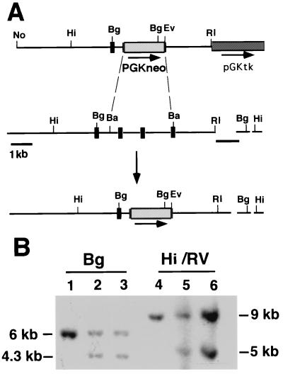
Figure 1.
(A) Disruption of smad2 through homologous recombination in embryonic stem (ES) cells. The targeting vector psmad2neo deletes the carboxyl-terminal portion of smad2. The top line is the targeting vector, the middle the genomic locus before recombination, and the bottom the locus after recombination. 5′ is on the left. The size and position of exons are not drawn to scale. The arrow represents the direction of neo transcription, the dashed lines the portion deleted, and the short black lines the 3′ flanking and 5′ internal probes used to detect homologous recombination. (B) Southern blot of ES DNAs. The 3′ flanking probe recognizes a 6-kb BglII fragment from the wild-type allele; this fragment is reduced to 4.3 kb in the targeted allele because of the presence of a BglII site on the neo gene. Correct targeting was confirmed with a HindIII/EcoRV digest, which releases a 9-kb band from the wild type and a 5-kb band from the targeted allele. The 5′ internal probe was used to further confirm correct targeting (data not shown). Both correctly targeted clones are shown. Hi, HindIII; No, NotI; Bg, BglII; Ba, BamHI; RI, EcoRI; and RV, EcoRV.

