Fig 2.

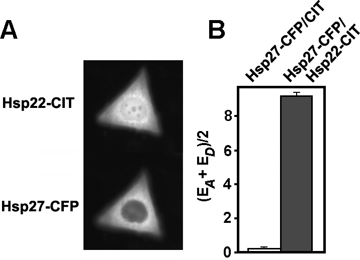
Interaction of Hsp22 and Hsp27 in estrogen receptor–positive (ER+) MCF-7 cells in vivo. Cells grown in complete medium were doubly transfected to express citrine (CIT) and cyan (CFP) fluorescent protein fusion proteins (Hsp27-CFP and Hsp22-CIT). (A) Fluorescence image of a representative cell coexpressing both sHSP fusion proteins. Expressed Hsp22-CIT was located in both the cytoplasm and nucleus, whereas expressed Hsp27-CFP was mainly located in the cytoplasm. (B) qFRET data collected from 69 doubly-transfected cells (gray bar) resulted in an average fluorescence resonance energy transfer efficiency (AAFE) value that was significantly different (P < 0.01) from the AAFE value collected from 63 control cells coexpressing Hsp27-CFP and CIT (white bar). Error bars represent standard error
