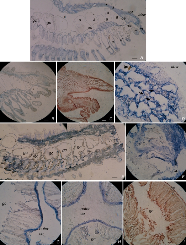
Fig 4.
Tracing coelomocytes in tissues of normal and amputated sea star arms. Longitudinal sections of (A–D) normal or (E–I) amputated arms, labelled with (A, D, E–H) anti-toposome McAb, (B) Domangk dye, or (C, I) hematoxylin dyes. The main recognizable anatomical features are: abw, aboral body wall; a, ampulla; ce, coeleomic epithelium, d, dermis; ep, epidermis; gc, gastric caecae; pc, perivisceral coelom; tf, tube feet; to, terminal ossicle; tt, terminal tentacle; rnc, radial nerve cord. On the right side (E) a line indicates the amputation plan. (D) Enlargement of the terminal ossicle shown in (A); arrowhead: red amoebocyte; arrows: white amoebocytes dispersed in the connective stroma of the skeletal plate. (F,G) Enlargements of the truncated stump and coelomic epithelium respectively, shown at lower magnification (E). Bar = 25 μm

