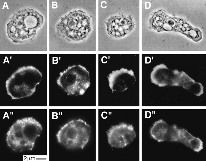
Figure 10.
Immunofluorescence localization of p35 and p40 in Acanthamoeba castellani. Cells fixed with 1% formaldehyde in 97.5% ice-cold methanol were stained with affinity-purified rabbit antibodies against either p35 (A and B) or p40 (C and D). (A–D) Phase contrast microscopy. (A″–D″) Localization of p35 and p40 detected with cy3-conjugated goat anti–rabbit secondary antibodies. (A′–D′) Actin filament localization with BODIPY-conjugated phalloidin. Both p35 and p40 were present throughout the cytoplasm and enriched in the cell cortex especially in fibrous structures within microspikes (C″) or entirely within the cell (A″). (E′and F′) Confocal images of 0.8 μm sections of fixed amebas stained with antibodies against p40 (E′ and F′). (E and F) Corresponding phase images. The confocal images emphasize the enrichment of p40 in regions of the cell cortex and the fibrous character of the staining pattern.


