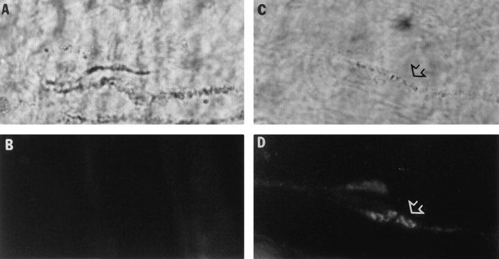
Figure 5.
Binding sites for AChE are regularly arrayed along the synaptic basal lamina. Empty frog basal lamina sheaths were incubated with either the G4/G2 (A and B), or A12 (C and D) quail AChE and the neuromuscular junctions localized histochemically (A and C). The quail enzyme was labeled by indirect immunofluorescence using mAb 1A2 (B and D). Only the A12 collagen-tailed AChE enzyme bound to the neuromuscular junction where it colocalized with the previously bound frog AChE.

