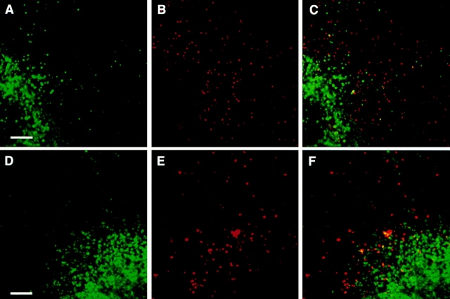
Figure 5.
K+ depletion inhibits ricin and endotubin colocalization. Cells were depleted of intracellular potassium and incubated with ricin as described in Fig. 4. After 2 min of uptake (A–C), endotubin staining (A) remains punctate in the periphery, but some diffuse perinuclear staining is evident. Ricin fluorescence (B) is distributed in a punctate pattern in the cell periphery, but there is no colocalization of endotubin and ricin (C). After 20 min of ricin uptake (D–F), endotubin staining is predominantly perinuclear (D). Ricin fluorescence remains punctate in the cell periphery (E), and there is no colocalization with endotubin staining (F). Bar equals 5 μm.

