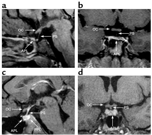
Figure 2.
Neuroimaging studies of the patient with the HESX1(I26T) mutation (a and b) and a normal subject (c and d). (a) Sagittal MRI scan of the patient homozygous for HESX1(I26T) at 21 years, showing a thin, continuous pituitary stalk (PS), a normal optic chiasm (OC), and a severely hypoplastic anterior pituitary lobe (APL). (b) Coronal MRI scan of the patient at 11 years, showing a hypoplastic anterior pituitary lobe and a normal optic chiasm, with a thin pituitary stalk that is not clearly visualized. (c) Normal sagittal MRI scan showing a normal anterior pituitary lobe, a normal pituitary stalk, a normally sited PPL, and a normal optic chiasm. (d) Normal coronal MRI scan showing a normal anterior pituitary lobe and a normal optic chiasm with a normal pituitary stalk.

