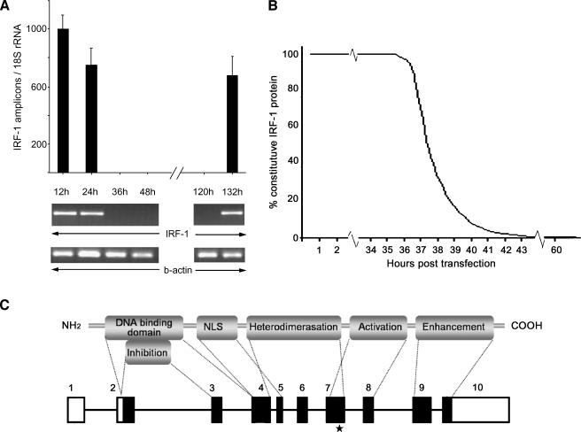
Figure 1.
Small interfering RNA for the transcriptional inhibition of IRF-1. (A) The IRF-1 transcriptional expression as assessed by real-time PCR. At 36 hours posttransfection, no IRF-1 mRNA was detected. IRF-1 expression was restored 136 hours posttransfection. The transcriptional expression of β-actin (PCR) and 18S rRNA (real-time PCR) were not disturbed by the process. The THP-1 cells treated with transfection reagent alone or non-silencing scrambled siRNA revealed no change in IRF-1 expression. (B) A mathematical approach of the degradation rate of the constitutive IRF-1 protein levels posttransfection. At 36 hours after the addition of the siRNA, the transcription of IRF-1 was inhibited. The already formed IRF-1 protein levels (constitutive) would drop by 50% every 30 minutes (half-life). We estimated that, at 42 hours posttransfection, the IRF-1 protein levels would be less than 0.1% of the constitutive. To further minimize the margin of error, we set the LPS induction at 52 hours and the harvesting time at 60 hours posttransfection. (C) Schematic representation of the IRF-1 protein's functional domains and the mRNA. The known functional domains of the IRF-1 protein are depicted and correlated with the genomic arrangement of the full-length transcript. The black boxes represent the translated areas. The asterisk indicates the complementary site of the siRNA, which is outside the conserved region shared by the IRF family members.

