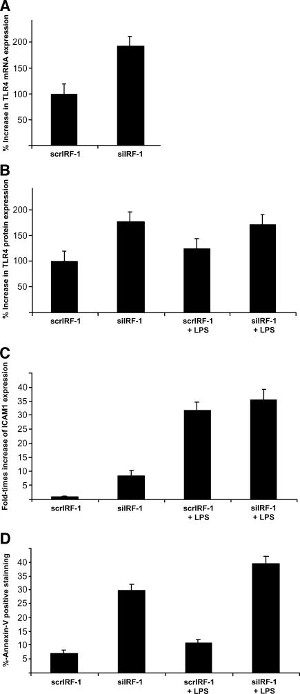
Figure 2.
siIRF-1 THP-1 cells present elevated levels of TLR-4 and ICAM.1 protein levels as well as increased Annexin-V+ staining. (A) The siIRF-1 THP-1 cells presented a 90 ± 10% increase of the TLR-4 mRNA levels when compared to the scrIRF-1 THP-1 cells (P < .01). (B) The siIRF-1 THP-1 cells presented a 77 ± 8% increase of the TLR-4 protein levels when compared to the scrIRF-1 THP-1 cells (P < .01). The scrIRF-1 THP-1 cells treated with LPS presented elevated TLR-4 protein levels by 25 ± 3% in comparison with the untreated cells. The siIRF-1 THP-1 cells challenged with LPS showed no differences in the TLR-4 protein levels when compared to the untreated siIRF-1 THP-1 cells. Finally, the LPS-treated siIRF-1 THP-1 cells presented a 46 ± 8% increase in the TLR-4 protein levels in contrast to the LPS-triggered scrIRF-1 THP-1 cells. (C) The siIRF-1 THP-1 cells presented an 8.35-fold increase in their ICAM.1 protein levels, in contrast to the scrIRF-1 THP-1 cells (P < .01). The scrIRF-1 THP-1 cells treated with LPS presented a 31.7-fold increase in ICAM.1 protein levels in comparison with the untreated cells (P < .01). The siIRF-1 THP-1 cells challenged with LPS revealed a 27.3-fold increase in ICAM.1 levels when compared to the untreated siIRF-1 THP-1 cells (P < .01). Finally, the LPS-treated siIRF-1 THP-1 cells presented a 3.98-fold increase in ICAM.1 levels in contrast to the LPS-triggered scrIRF-1 THP-1 cells (P < .01). (D) The constitutive levels of apoptosis (Annexin-V+) in the scrIRF-1 THP-1 cells were calculated at 7 ± 2% of the total population. The siIRF-1 THP-1 cells presented a significant increase in apoptosis (4.25-fold) with the Annexin-V+ positive cells reaching 29 ± 3% of the total population, indicating that IRF-1 interference resulted in a 23% increase in cellular apoptotic levels (P < .01). The scrIRF-1 THP-1 cells treated with LPS presented a 0.55-fold increase in Annexin-V+ staining (10.85 ± 2%) in comparison with the untreated cells. The siIRF-1 THP-1 cells challenged with LPS (39.5 ± 4%), revealed a 1.4-fold increase in the apoptotic levels when compared to the untreated siIRF-1 THP-1 cells (P < .05). Finally, the LPS-treated siIRF-1 THP-1 cells presented a 3.6-fold increase in Annexin-V+ staining in contrast to the LPS-triggered scrIRF-1 THP-1 cells (P < .01).

