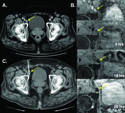
Figure 3.
Benign right external iliac node. (A) Axial contrast-enhanced computed tomographic (CT) image shows an enlarged right external iliac node. (B) Sequential images show progressive loss of signal following ferumoxytol administration. (C) CT-guided biopsy showing biopsy needle within the enlarged lymph node.

