
Figure 3.
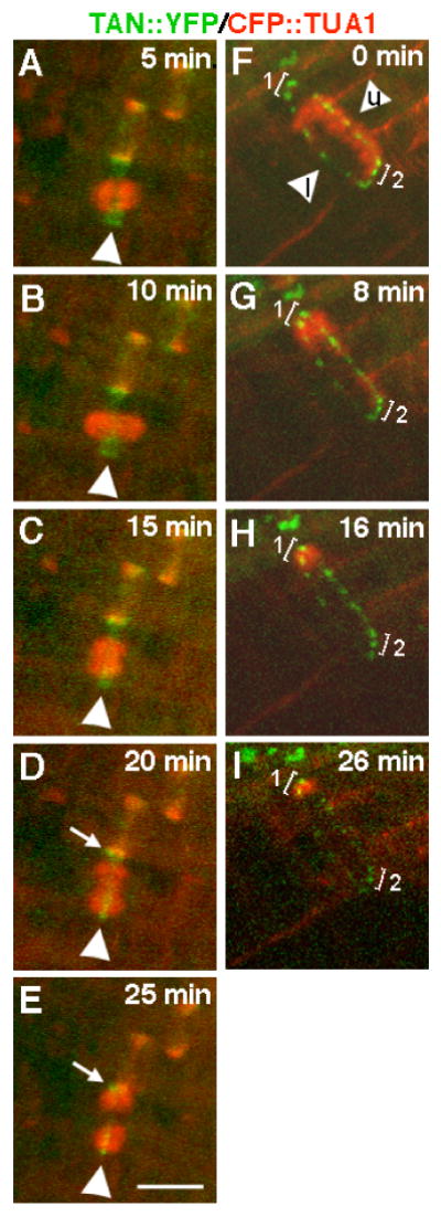
Timelapse analysis of changes in the appearance of AtTAN::YFP rings (green) during cell division with CFP::TUA1 shown in red. A-F, The metaphase cell shown at time zero in Figure 2E-H was subsequently imaged every five minutes as indicated. Arrowheads point to an AtTAN::YFP ring in a cell with a metaphase spindle (A), an elongated spindle (B), a newly initiated phragmoplast (D), an expanded phragmoplast (D), and a more expanded phragmoplast (E). Arrows in D and E point to the edge of a broad, PPB-associated AtTAN::YFP ring in the adjacent cell. F-I, A cell completing cytokinesis was imaged every 8-10 minutes as indicated. In F, a highly punctate, but complete, AtTAN::YFP ring encircles a cell with a phragmoplast that has expanded to the upper face of the cell (arrowhead marked “u”) and has already disassembled at the lower face (arrowhead marked “l”; note that AtTAN::YFP puncta in bracketed areas 1 and 2 are equally bright at time zero). Subsequently, the phragmoplast completes its expansion into the corner marked by bracket 1 while disassembling elsewhere. By 8 minutes (G), the AtTAN::YFP ring has already begun to disintegrate at the lower face of the cell and by 26 minutes (I), has also disintegrated at the upper face and at the area marked by bracket 2 while persisting at the corner marked with bracket 1, where the phragmoplast has not yet disassembled. Scale bar = 10μm.