
Figure 4.
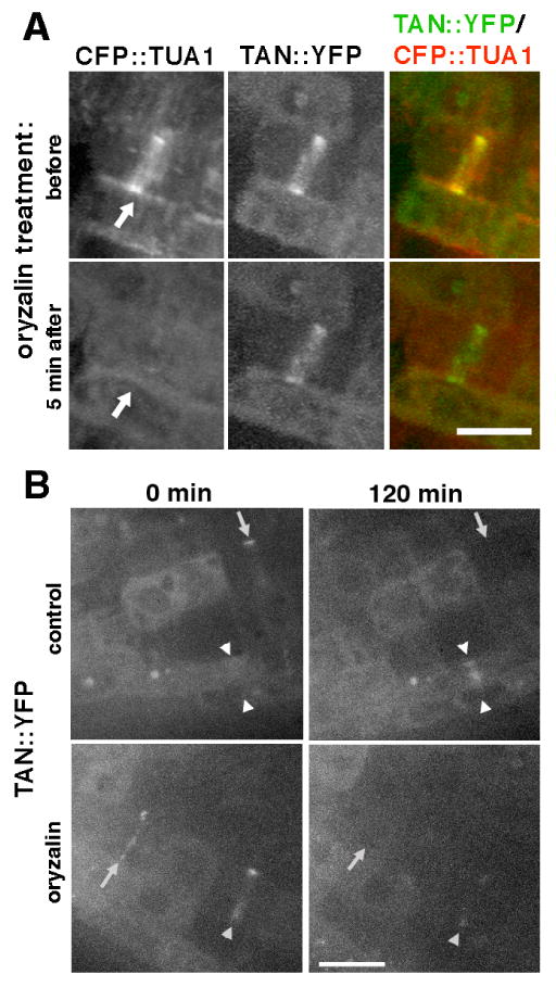
Analysis of the role of microtubules in AtTAN::YFP recruitment and retention at the division site (Z-projections of confocal stacks). A, CFP::TUA1 (monochrome in first column, red in third column) and AtTAN::YFP (monochrome in second column, green in third column) in a cell with a PPB (arrows) before (top) and 5 minutes after (bottom) perfusion of 5 μM oryzalin under the coverslip. The AtTAN::YFP ring persists after disassembly of PPB microtubules. B, AtTAN::YFP rings (monochrome only) observed in root tips treated with 5 μM oryzalin (bottom) or no oryzalin (top). Root tips were examined at 0 minutes (left) and again at 120 minutes (right). Comparison of 0 minute and 120 minute images for the same roots revealed rings that disappeared (arrows) or faded (single arrowhead) during the observation period in the presence or absence of oryzalin, but new rings (i.e., absent at 0 minutes but present at 120 minutes; double arrowheads) formed only in the absence of oryzalin. Scale bar = 10 μm.