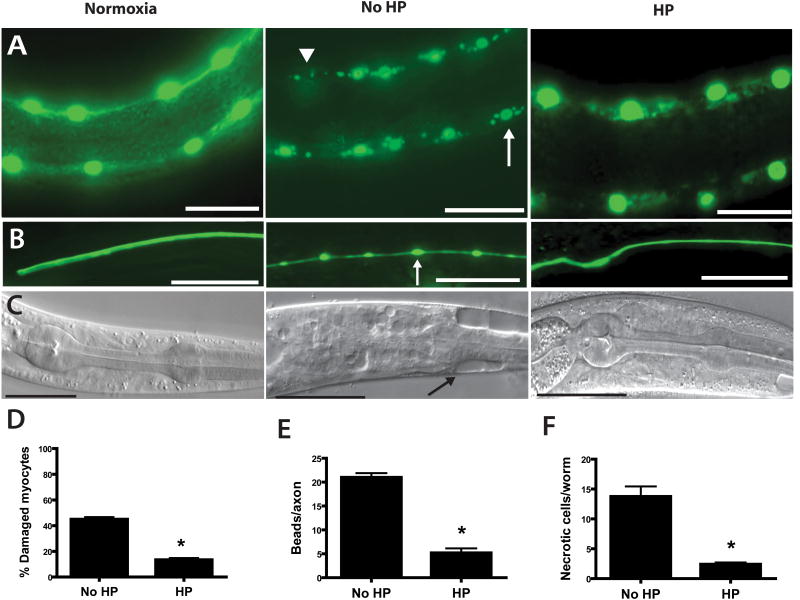
Figure 2. Hypoxia induced pathological cell defects and cell death blocked by HP.
L1 larvae underwent no hypoxic incubation (Normoxia) or a 20 hr hypoxic incubation that was preceded by 4 hr HP (HP) or normoxic preconditioning (No HP). After a 24 hr recovery, surviving animals were scored for cell pathological defects. Scale bars = 20μm.
(A,D) Muscle nuclear pathology reduced by HP. Muscle nuclei were visualized by nuclear-localized GFP driven by a muscle-specific promoter – Pmyo-3. Hypoxia produces damaged myocytes seen as nuclear fragmentation (arrow) and nuclear loss (arrowhead) [5] that is reduced by HP (* - p < 0.01 vs No HP, n=29 animals no HP, 44 HP).
(B,E) Axonal pathology reduced by HP. Touch sensory neurons were visualized by GFP driven by a touch neuron-specific promoter – Pmec-4. Hypoxia produces axonal beading pathology (arrow) [5] that is reduced by HP (* - p < 0.01 vs No HP, mean ± sem, n=124 axons no HP, 116 HP).
(C,F) Necrotic cell death reduced by HP. Hypoxia produces necrotic cell death (arrow) [5] that is reduced by HP (* - p < 0.01 vs No HP, n=29 animals no HP, 44 HP).

