
Figure 2. γHV-68 vMAP Encodes a Mitochondrial Protein.
(A) Identification of vMAP protein. (Left panel) γHV-68-infected (lane 2 and 3) or mock-infected NIH3T3 cells (lane 1) were harvested at 16 h post-infection. Post-centrifuged lysates in CHAPS buffer were precipitated with pre-immune serum (lane 2) or anti-vMAP serum (lanes 1 and 3), followed by immunoblotting (IB) with anti-vMAP serum. The arrowhead indicates vMAP. (Right panel) Subcellular fractionation of vMAP in 293T cells at 36 h post-transfection, as shown by immunoblotting with antibodies to vMAP, COX4, or actin. W, whole cell lysate; C, cytosolic; HM, mitochondrion-enriched heavy membrane.
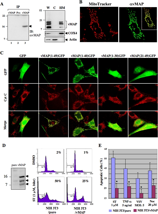
(B) vMAP localizes to the mitochondrion. Post-transfection with pcDNA5-vMAP vector, COS-1 cells were stained with MitoTracker and fixed for anti-vMAP immunostaining. The box inside of the right panel shows the merged image of MitoTracker and anti-vMAP immunostaining.
(C) Identification of the vMAP MTS. Either GFP alone or various vMAP N-terminal sequences fused to GFP were expressed in NIH3T3 cells. Mitochondria were stained with anti–cytochrome c antibody (red). Pictures represent more than 85% of cells analyzed by fluorescent microscopy.
(D) vMAP inhibits apoptosis. NIH3T3/puro and NIH3T3/vMAP cells were treated with DMSO or ST (1 μM) for 16 h and stained with PI, followed by flow cytometry analysis. Data are from one of three replicate experiments. vMAP (indicated by arrows) expression was shown by immunoblotting on the left.
(E) vMAP inhibits apoptosis initiated by various apoptogenic stimuli. NIH3T3/puro and NIH3T3/vMAP cells were treated with various agents (ST, TNF-α/cycloheximide, vesicular stomatitis [VSV] infection for 16 h, or nocodazole [Noc] for 36 h), and sub-G1 cells were quantified as described in (D). Data represent result of three independent experiments and error bars indicate standard deviation with (*) p < 0.05 relative to control (puro) as calculated by Student's t-test.