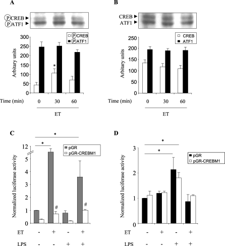
Figure 3. ET Induces CREB Activation.
AMs were incubated for 2 h with ET (1 μg/ml) and were then stimulated by LPS (50 ng/ml). After incubation for the time periods indicated, total cell proteins were extracted.
(A and B) Western blot analyzes were then performed using phospho-CREB (ser 133) (A) or CREB (B) antibodies. The figure also shows the phosphorylation (A) and expression (B) levels of the activating transcription factor-1 (ATF-1). Corresponding quantifications were carried out using densitometry and were expressed as arbitrary units (representative of three separate experiments).
(C) CHO cells were transfected with CREB ([CRE]4-Luc) reporter plasmid and/or dominant-negative CREB construct (pGR-CREBM1) or vehicle (pGR) for 24 h, pretreated for 1 h with ET (1 μg/ml) and stimulated with LPS (50 ng/ml) for an additional 24 h.
(D) CHO cells were transfected with the sPLA2-IIA-Luc reporter plasmid and/or pGR-CREBM1. After 24-h transfection, cells were pretreated for 1 h with ET (1 μg/ml) and were stimulated with LPS (50 ng/ml) for an additional 24 h. Cells were then lysed, and the luciferase activity was measured and normalized with β-galactosidase activity.
The data are the mean ± SEM and are representative of four separate experiments. An asterisk (*) indicates p < 0.05 ; a hash mark (#) indicates p < 0.05 compared to corresponding pGR controls.

