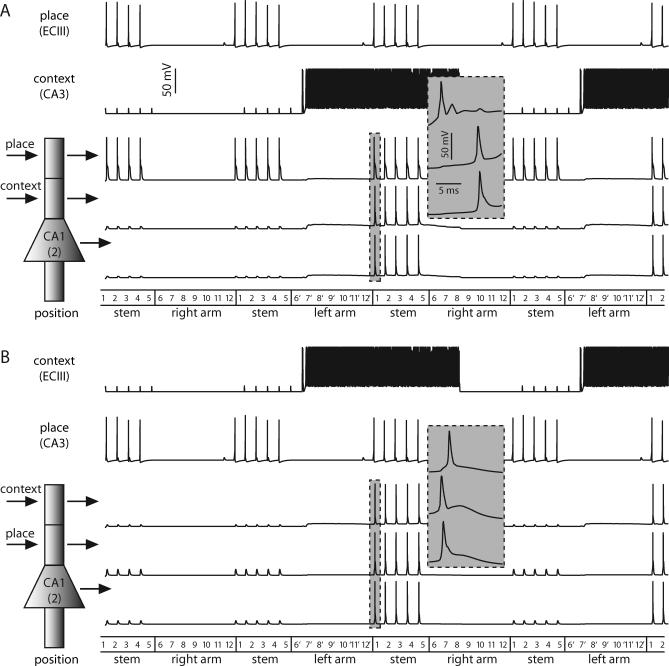
Figure 6. Gating in the Reduced Model of a CA1 Pyramidal Neuron.
(A) Shown are the cells representing position 2 of the maze. Here, the ECIII cell encodes place information and the CA3 cell represents temporal context. The CA1 cell only fires somatic spikes when the ECIII and CA3 inputs are coincident. As the rat enters the stem from the right arm, the subthreshold responses in the proximal apical dendrites and soma correspond to dendritic spikes that fail as they propagate forward. The gray inset shows the first set of CA1 spikes on an expanded time scale.
(B) Same as above except the ECIII cell represents temporal context and the CA3 cell encodes raw place information. Although the somatic action potential profiles in (A) and (B) are roughly identical; in this case, the spike is initiated in the proximal apical dendritic compartment and propagates forward to the soma and backward to the apical tuft, as can be seen in the gray inset.

