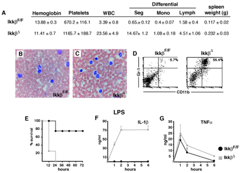
Figure 3. IkkβΔ mice develop granulocytosis and show massively increased circulating IL-1β after endotoxin exposure.

(A) Complete and differential blood counts and spleen weights of IkkβF/F and IkkβΔ mice. Data are average values for 5 mice of each genotype examined two weeks after a single poly-(I:C) injection. (B, C) Blood smears stained with Wright-Giemsa and (D) number of CD11b+/Gr-1+ cells in spleens of of IkkβF/F and IkkβΔ mice analyzed two weeks after poly-(I:C) injection. (E) Survival after endotoxin injection (20 mg/kg, E. coli O111:B4) of IkkβF/F (black) and IkkβΔ mice (grey), (n = 4–6). (F) IL-1β and (G) TNF-α plasma levels 1, 2 and 6 hrs after LPS administration.
