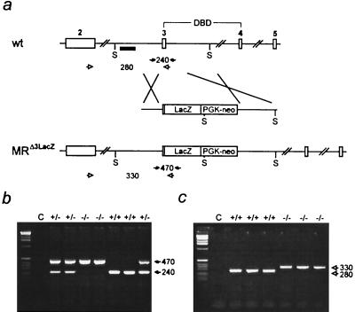
Figure 1.
Inactivation of the mouse MR gene by gene targeting. (a) Targeting strategy. (Top) Part of the MR gene with exons 2, 3, 4, and 5. Exons 3 and 4 encode the two zinc fingers of the DNA binding domain (DBD). S indicates SpeI restriction sites, and the small black box indicates the 5′ probe used for Southern blot analysis. (Middle) Targeting construct. LacZ and PGK neo indicate the β-galactosidase gene and the neomycin-resistance gene driven by the phosphoglycerate kinase promoter. (Bottom) Targeted MR locus. (b) Genotyping by PCR of genomic tail DNA by using specific primers (filled arrows in a). Numbers between the arrows indicate the size of the amplified fragments in bp. C, water control. (c) Reverse transcription–PCR analysis of cDNA derived from total kidney RNA of wild-type (+/+) and MR-deficient (−/−) mice by using exon- and LacZ-specific primers (open arrows in a) demonstrates the absence of the 280-bp wild-type-specific band in MR −/− mice. Instead, only the 330-bp band specific for the MRΔ3-LacZ fusion mRNA is present.

