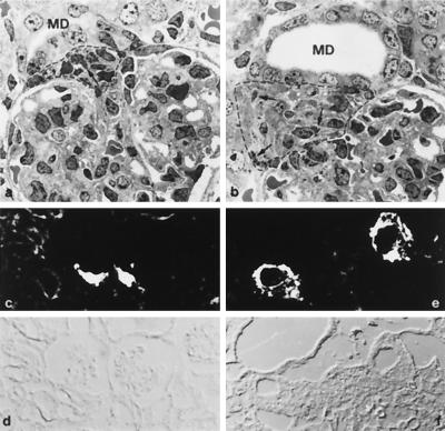
Figure 3.
Histological and immunocytochemical analysis of the kidney. Transmission electron micrographs of glomeruli and vascular pole region of wild-type (a) and MR −/− mice (b) from day 8 after birth. In MR −/− mice, the macula densa segment of the distal tubule (MD) was enlarged. The extraglomerular mesangium (encircled by a dotted line) showed prominent hyperplasia with granules (arrows in a and b) in almost every cell. Renin immunocytochemistry (c and e) and corresponding phase contrast pictures (d and f) of 8-day-old mice. In MR −/− mice, (e) staining for renin at the vascular pole region is much more prominent than in wild-type mice (c).

