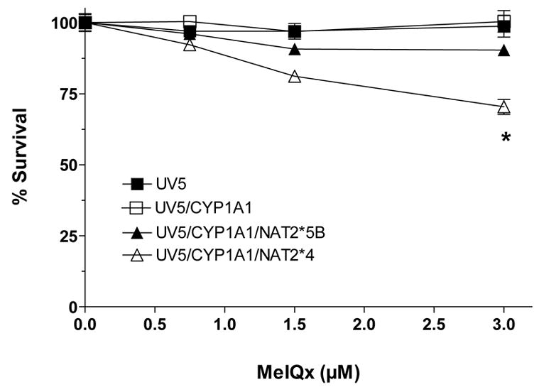
Figure 3.

MeIQx-induced cytotoxicity in UV5 CHO cell lines. Percent survival on the ordinate is plotted versus MeIQx treatment concentration on the abscissa. Each data point represents Mean ± S.E.M. for three experiments. MeIQx-induced cytotoxicity was significantly greater (p<0.05) in CYP1A1/NAT2*4- than CYP1A1/NAT2*5B-transfected CHO cells at 3 μM.
