
Figure 5.
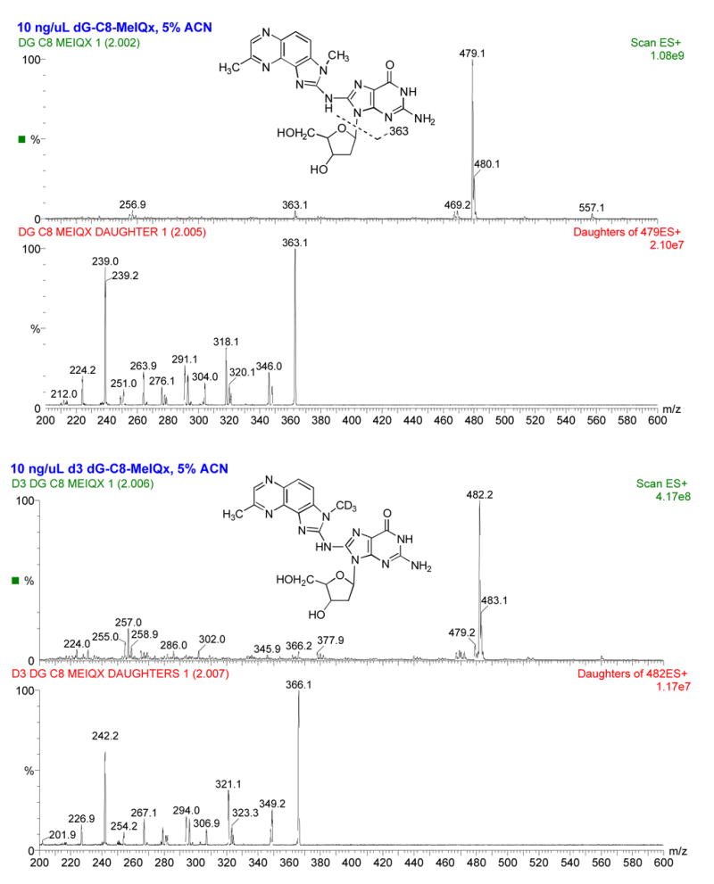
Structure and electrospray ionization spectra of dG-C8-MeIQx (top two panels) and dG-C8-MeIQx-D3 (bottom two panels) showing [M+H]+ = 479 and 482, respectively. Collision induced dissociation fragmentation of dG-C8-MeIQx and d3 dG-C8-MeIQx at −50 V collision energy shows fragmentation of the MeIQx and guanosine moieties dominated by the major fragment produced from loss of deoxyribose: the aglycone ion of m/z 363 or 366 for dG-C8-MeIQx and dG-C8-MeIQx-D3, respectively. The collision energy was −25 V for MRM transitions used during quantitation.