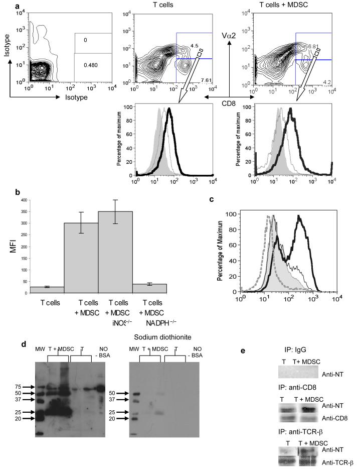
Figure 4. Role of MDSC in the nitration of tyrosine in a CD8+ T cell.
(a) Adoptive transfer and immunization was performed as described in Figs 1 and S1. Eight days later LNs were isolated and labeled with anti-CD8, anti-Vα2, and anti-NT antibodies. The level of NT expression was evaluated within population of donors (CD8+Vα2+) (bold line) and recipient (CD8+Vα2−) (thin line). Shaded areas show staining with isotype control IgG.
(b) Splenocytes from OT-1 mice were cultured with control or specific peptides for 72 h in the presence of MDSC (at 3:1 ratio) from wild-type, NADPH−/−(gp91phox−/−) and iNOS−/− EL-4 tumor-bearing mice. After that time cells were collected and the levels of NT in CD8+ T cells were evaluated by flow cytometry. Mean ± SD of MFI from three experiments are shown. Not shown MFI for samples with control peptides, which were below 30 in all experiments.
(c) The level of NT in CD8+ OT-1 T cells. Cells were treated as described in Fig. 4b. Shaded area − splenocytes alone, bold line splenocytes + MDSC; dashed line 1 mM SOD was added; thin line − 1 mM L-NMMA was added. CD8+ cells were gated and NT expression was measured within this cell population. Not shown MFI for samples with control peptides, which were below 30 in all experiments.
(d) Splenocytes from OT-1 mice were cultured for 48 h with specific peptide in the presence of MDSC (at 3:1 ratio). Whole cell lysates were prepared and were subjected to 10%SDS-PAGE electrophoresis and probed with anti-NT antibody. Nitrated BSA was used as a positive control for NT staining. Bands were visualized using ECL. Chemical reduction of nitrotyrosine was done using sodium dithionite. The membrane was washed and probed with anti-NT antibody. Each experiment was performed in duplicates (shown).
(e) Splenocytes cells from OT-1 mice were treated as described above. Whole cell lysates were prepared and CD8 and TCRβ were immunoprecipitated using specific antibodies as described in Methods. Rabbit IgG was used in control experiments. The proteins were subjected to 10%SDS-PAGE electrophoresis and probed with anti-NT antibody. To verify loading, the same blots were stripped and re-probed with anti-CD8 or anti-TCRβ antibodies.

