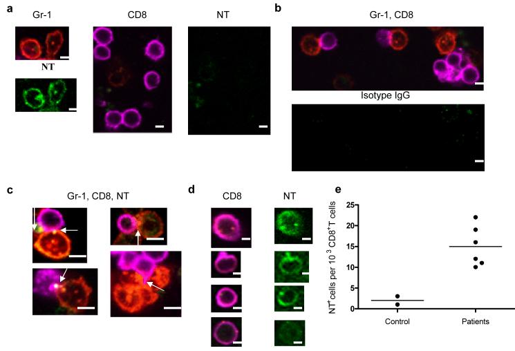
Figure 5. Interaction between MDSC and CD8+ T cells.
(a–d) Splenocytes from OT-1 mice were cultured with specific peptide and MDSC (at 3:1 ratio). The cells were labeled with anti-Gr-1-PE (red), anti-CD8-Alexa 647 (magenta), anti-NT-Alexa 488 (green), or isotype-Alexa 488 (green) at different time points and visualized under a confocal microscopy. Scale bar = 10 μm. (a) MDSC and CD8 T cells separate; (b–c) 5 h co-culture. Arrows denote positive staining for NT localized at contacts between MDSC and CD8+ T cells. (d) 48 h culture.
(e) LN from individuals with breast and head and neck cancers were collected during tumor resection as part of routine pathological examination. LNs without presence of tumor were further evaluated (see Fig. S5). The number of NT positive cells per 103 CD8+ T cells are shown.

