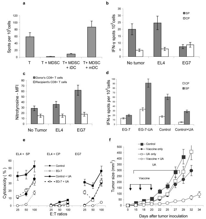
Figure 6. MDSC induced CD8+ T-cell tolerance in tumor-bearing mice.
(a) Mice were treated as described in Fig. S1. Ten days after immunization LN cells were re-stimulated in triplicates with control or specific peptide or with bone marrow derived immature (iDC) or LPS activated matured (mDC) DCs. DCs were added at 1:10 ratio. The number of IFN-γ producing cells was measured in ELISPOT assay. Mean ± st. dev. from two experiments are shown. The values of stimulation with control peptide were subtracted from the values of stimulation with specific peptide.
(b–e). C57BL/6 mice were inoculated subcutaneously with 5×10⁵ of EL-4 or EG-7 tumor cells. Seven days after inoculation each mouse had been injected intravenously with 5×10⁶ OT-1 T-cells. Seven days later mice were sacrificed and LN cells used in further experiments. (b) LN cells were stimulated with specific (SP) (SIINFEKL) or control (CP) (RAHYNIVTF) peptides and analyzed in IFN-γ ELISPOT assay. The number of spots were calculated per 105 LN cells. Each group included 3 mice. The differences between SP and CP were statistically significant (p<0.05) from control and EL-4 tumor-bearing mice. (c) Splenocytes were labeled with anti-CD8, anti-Vα2' and anti-nitrotyrosine antibodies. The level of NT expression was evaluated within the populations of donors (CD8+Vα2+) and recipient (CD8+Vα2−) cells. MFI-mean fluorescence intensity. Results of the two performed experiments are shown. The differences between NT levels in donor's cells from EG-7 mice and EL-4 or control mice were statistically significant. (d) EG-7 tumor-bearing mice were treated with daily i.p. injections of UA (20 mg/100 μl PBS) starting from the day of T-cell transfer. LN cells were collected 7 days later and analyzed in IFN-γ ELISPOT assay as described in Fig. 6b. (e) Splenocytes from EG-7 tumor-bearing mice described above were stimulated ex vivo for 7 days with SIINFEKL peptide 10 μg/ml, in the presence of IL-15 (10 ng/ml) and IL-21 (10 ng/ml) and used as effectors in a standard 6-h 51Cr-release CTL assay. Target cells were EL-4 cells pulsed with 10 μg/ml of either specific (SIINFEKL) or control (RAHYNIVTF) peptides. In addition, EG7 cells were also used as targets. Different effector:target cell ratios were used. Each experiment was performed in duplicates and included three mice per group. Mean ± st. dev. are shown.
(f) MC38 tumor was established in C57BL/6 mice by subcutaneous injection of 2×10⁵ tumor cells. On day 10 after tumor inoculation when tumors became palpable mice were split into four groups with equal tumor size. Each group included 5-6 mice. Mice in treatment group (vaccine + UA) were injected s.c with 5×10⁵ DCs infected recombinant adenovirus containing mouse wild-type p53 gene as previously described29. Immunizations were repeated twice with 5-6 day interval. Treatment with UA (20 mg/100 μl PBS) was started three days after the first immunization and was continued for two weeks. Control groups included mice treated with UA alone (UA only), vaccine alone (vaccine only), or untreated (control). Tumor size was measured using calipers and presented as the result of multiplication of two longest dimensions. Mean ± st. dev. are shown.

