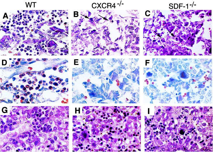
Figure 3.
Defects in B-lymphopoiesis and myelopoiesis in CXCR4−/− and SDF-1−/− mice. (A–C) Sections of femur bone marrow stained with H&E. Arrows indicate erythroid and megakaryocytic cells. (D–F) Sections of bone marrow stained for chloroacetate esterase activity. (G–I) Sections of liver stained with H&E.

