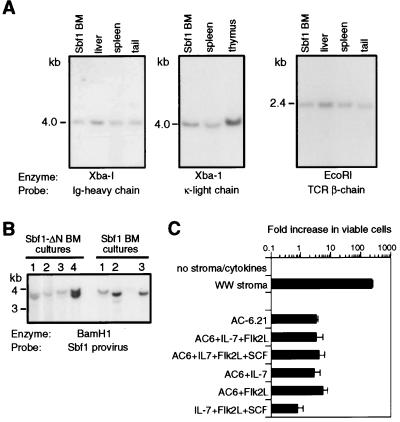
Figure 5.
Proviral integration, antigen receptor gene configurations, and growth requirements of Sbf1-expressing cells. (A) Southern blot analysis was performed by using enzymes and probes indicated beneath the respective panels. (B) DNA was isolated from bone marrow cultures initiated by either Sbf1 or Sbf1ΔN retroviruses and subjected to Southern blot analysis using a probe specific for the pSRαMSV-Sbf1 vector to assess the configurations of retroviral integration sites. Each of the several detected bands represent clonal expansions of cells with integrated Sbf1 proviruses. (C) Growth responses of Sbf1-expressing cells are displayed as the fold increase in viable cells after 5 days in media containing the indicated cytokines with or without bone marrow-derived stroma or the AC-6.21 stromal cell line.

