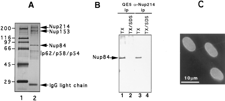
Figure 1.
(A) Nucleoporins immunoadsorbed from rat liver nuclear envelopes using QE5-Sepharose beads (lane 2). The material shown is derived from 1 to 2 g of rat liver and is revealed by silver staining. Approximately 50– 100× this quantity was used for limited amino acid sequence analysis of Nup84. Molecular weight markers (indicated in kD) are shown in lane 1. (B) Western blot analysis of QE5 and antiNup214 immunoprecipitates of NRK cells using an antipeptide antibody against Nup84. Immunoprecipitates were washed either in a low stringency buffer containing 0.5% Triton X-100 (TX, lanes 1 and 3) that preserves the Nup214–Nup84 interaction, or in a high stringency buffer containing 0.5% Triton X-100 plus 0.1% SDS (TX/SDS, lanes 2 and 4). This latter buffer breaks the interaction between Nup214 and Nup84 (Panté et al., 1994). (C) Indirect immunofluorescence labeling of NRK cells using the anti-peptide antibody against Nup84. The nuclear envelope is clearly labeled.

