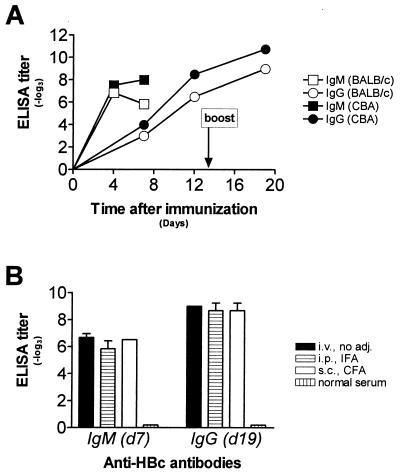
Figure 1.
Antibody response to HBcAg in different mouse strains and after various immunization procedures. (A) BALB/c (open symbols) and CBA (closed symbols) mice were immunized with HBcAg on day 0 and boosted with the same amount on day 13. IgM (squares) and IgG titers (circles) were determined in an ELISA assays on plates coated with HBcAg. Each data point represents the mean of three mice. (B) BALB/c mice were immunized with HBcAg as mentioned above either i.v. (black bars), i.p. in incomplete Freund’s adjuvant (horizontally striped bars), or s.c. in complete Freund’s adjuvant (open bars). Primary IgM titers on day 7 and secondary IgG titers on day 19 were determined by ELISA. Titers are indicated as −log3 of 20-fold prediluted sera. Each bar represents the mean of two to three animals; experiments were repeated twice.

