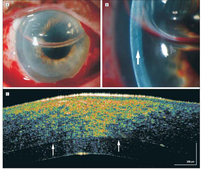
Figure 5.
Descemet membrane stripping endokeratoplasty. A, On postoperative day 1, 50% air bubble fill in the anterior chamber helps with adherence of the transplant. B, Magnified slitlamp view of the high-reflectivity donor-host interface (arrow). C, The interface between the recipient and donor corneas is marked by arrows. High-speed, ultra–high-resolution optical coherence tomography shows the strongest light signal approaching the epithelial surface. In addition to the proximity to the strongest light signal the patient's own corneal tissue is edematous and hyperreflective secondary to Fuchs endothelial dystrophy. The inner donor stromal–Descemet membrane–endothelial transplant is clearly seen as hyporeflective behind the hyperreflective donor-host interface. The donor and original corneas are seen in full adherence.

