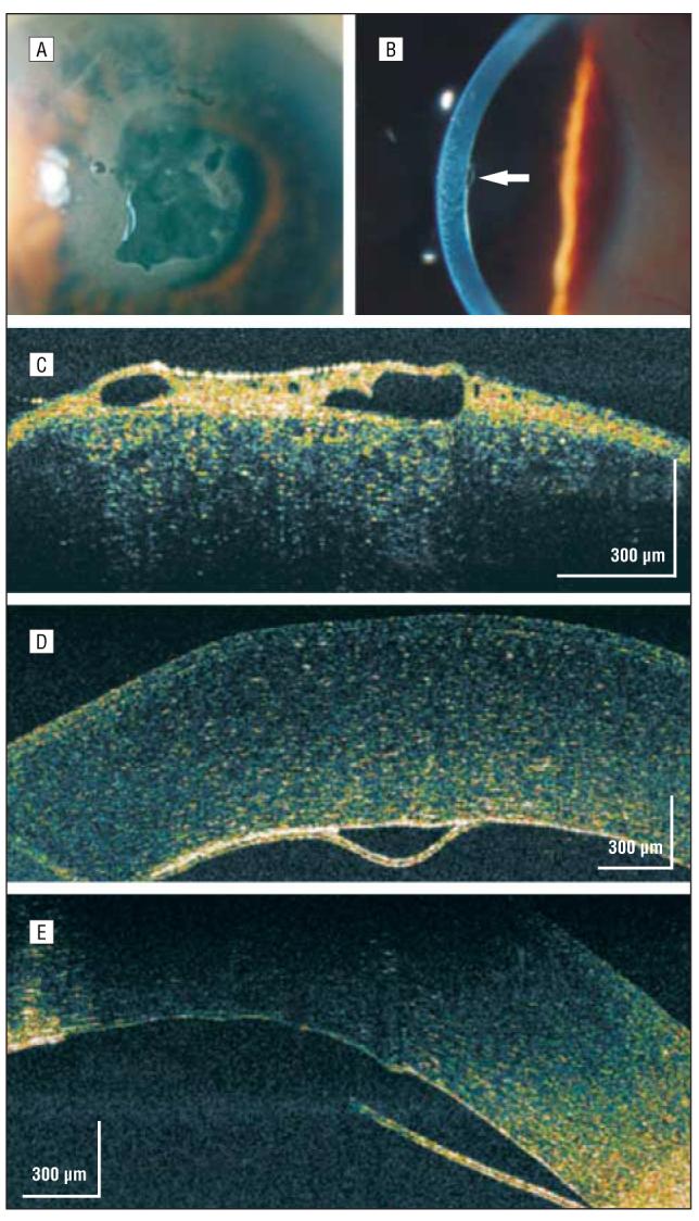
Figure 9.

Descemet membrane detachment. A, Persistent central bullous corneal edema of unestablished etiology 1 month after phacoemulsification. B, On high-magnification slitlamp photography, a small nasal Descemet membrane detachment was noted (arrow). C, High-speed, ultra–high-resolution optical coherence tomography (hsUHR-OCT) shows a thickened epithelium with large hyporeflective corneal epithelial bullae. D, An hsUHR-OCT image highlights the small Descemet membrane separation adjacent to the missing Descemet membrane to the right. E, A more central view of the Descemet membrane detachment with the adjacent missing Descemet membrane consistent with the overlying central area of bullous epithelium (not shown).
