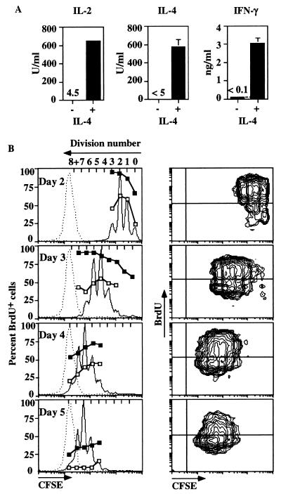
Figure 1.
IL-4 production and asynchronous division of stimulated T cells. (A) T cells were stimulated in primary culture with immobilized anti-CD3 and IL-2 with or without IL-4. After 4 days, activated cells were washed and restimulated with anti-CD3 (10 μg/ml) for 24 h. Supernatants were assayed for IL-2, IL-4, and IFN-γ. (B) CFSE profiles and BrdU labeling of dividing T cells at different times. CFSE-labeled T cells were stimulated with anti-CD3, IL-2, and IL-4 and harvested on days 2–5 as indicated. Histograms of CFSE fluorescence are shown with autofluorescence of nonlabeled cells overlayed as a dotted line (Left). (Right) BrdU incorporation by CFSE-labeled cells after a 9-h pulse (7 h for day 2). Contours (log density, 70%) are plotted with CFSE as abscissa and BrdU as ordinate. The percent BrdU+ cells in each division at each time is overlayed on the division histogram on the left and shows percent positive after either 1 h (open symbol) or 9 h (closed symbol).

