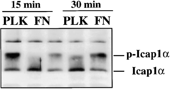
Figure 4.
Regulation of ICAP-1α phosphorylation during cell adhesion. UTA-6 cells were trypsinized and replated on plates coated with poly-l-lysine (PLK) (lanes 1 and 3) or fibronectin (FN) (lanes 2 and 4). Adherent cells were lysed in 0.5% NP-40 at t = 15 min (lanes 1 and 2), and t = 30 min (lanes 3 and 4) and the extent of ICAP-1α phosphorylation was determined on a Western blot using anti-ICAP-1 antibody.

