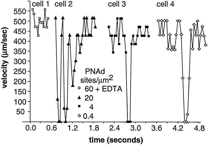
Figure 3.
The microkinetics of transient tethers on PNAd. Neutrophils were perfused at 1.5 dyn/cm2 on substrates with PNAd at the indicated density. Velocities of representative cells on each substrate are shown. Cell 1 in EDTA is shown to demonstrate the hydrodynamic velocity. Coordinates of cell centers were determined within one pixel accuracy (±0.9 μm) and the velocity between each frame was calculated for individual cells.

