
Figure 1.
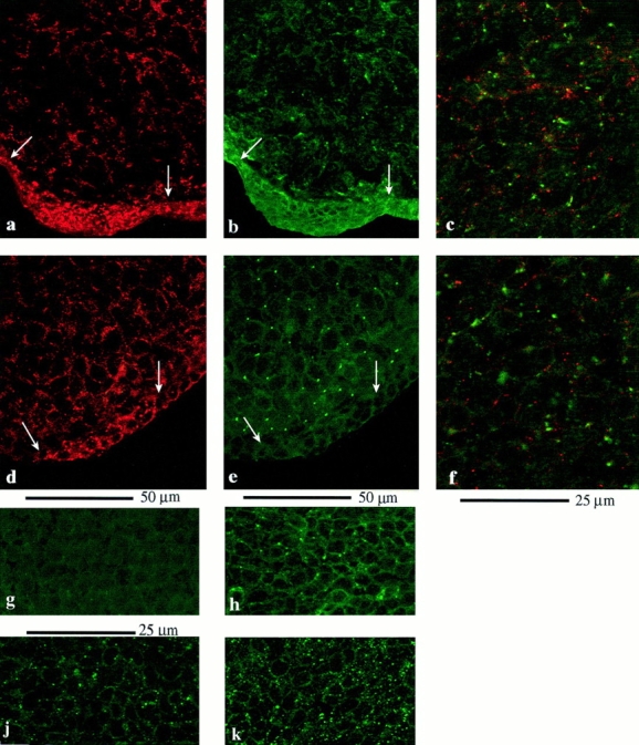
(a–h) Connexin staining in the intact limb bud. Sections through chick (a–c) and mouse (d–h) limb buds to show the pattern of gap junction protein expression. (a) Section through the tip of a chick limb bud stained for Cx43. Note dense expression of Cx43 between cells in the AER (between arrows). (b) Same section stained for Cx32. Occasional Cx32 gap junctions are present in the ridge ectoderm. Subapical mesenchyme cells show widespread expression of Cx32. (c) Region of the mesenchyme showing double labeling for Cx43 (red) and Cx32 (green). Note that although almost every cell expresses both connexin proteins, there is virtually no overlap between Cx32- and Cx43-containing plaques. (d–f) Equivalent sections through tip of mouse limb bud. Note that in the ectoderm, Cx43 (d) expression is restricted to the ridge itself and there is substantial expression in the mesenchyme, while Cx32 (e) is extremely rare in the ectoderm, although abundant in the mesenchyme. f shows both connexins. Again, there is no overlap between Cx43- (red) and Cx32- (green) containing gap junction plaques. (g and h) Cx32 staining by Des 5 is abolished by Des 5 peptide (g), but not by Gap 15 peptide (Cx43; h). j and k illustrate the lower Cx43 density (gap 15 antibodies) in chick posterior mesenchyme cultures maintained in the absence of FGF4 (j). Cx43 gap junction density is increased in posterior mesenchyme cultures maintained in FGF4 (k).