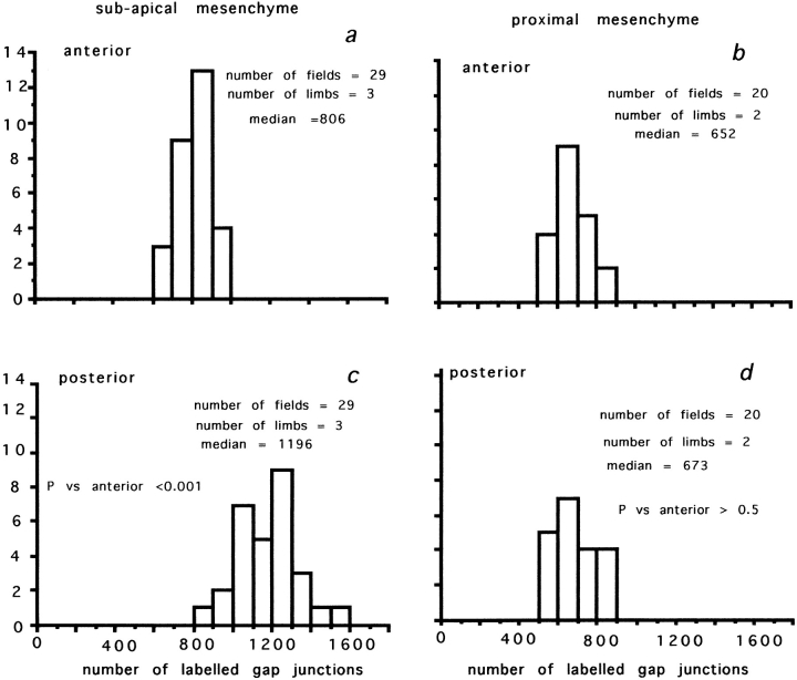
Figure 2.
The density of Cx43-labeled gap junctions in the chick limb bud at HH stage 19–20. (a and c) Comparison of frequency distributions for gap junctions counted between subapical mesenchyme cells below the AER. (a) Anterior; (c) posterior. Note that the density of gap junctions is significantly (P < 0.001) greater between posterior mesenchyme cells, the zone of polarizing activity. (b and d) Frequency distributions for the density of gap junctions between posterior and anterior mesenchyme cells in proximal regions (150 μm back from the AER). (b) Anterior; (d) posterior. Note no difference between posterior and anterior mesenchyme.

