
Figure 3.
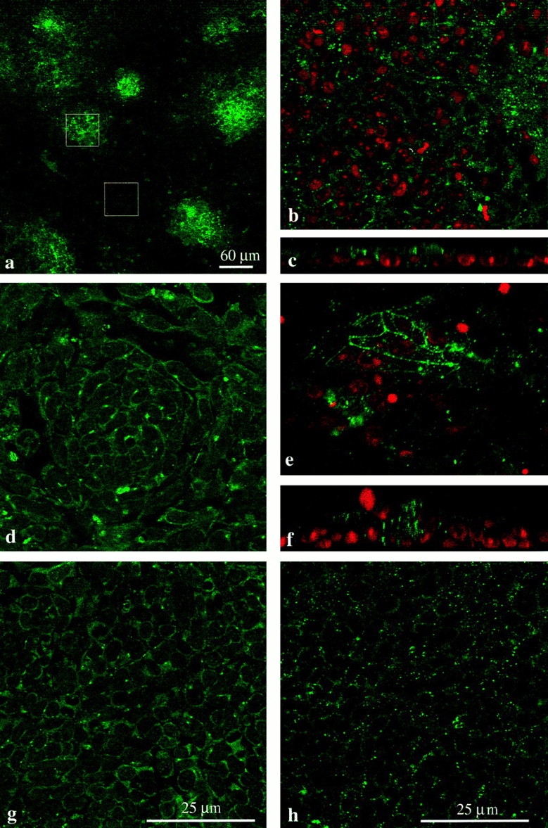
Connexin staining in micromass cultures. (a–c, e, and f) Chick mesenchyme; (d, g, and h) mouse mesenchyme. All images apart from a are at the same magnification. (a) Low power micrograph of chick mesenchyme culture stained for Cx43. The culture is made up of a large monolayer of undifferentiated cells interspersed with small condensations, which can be identified by the dense clustering of Cx43 staining. The box overlays show examples of regions chosen for counting. A 60 × 60–μm-sided square (3,600 μm2) either encloses a precartilage condensation or a region of the monolayer. (b) Example of region enclosed by 60-μm-sided square over an undifferentiated part of a chick mesenchyme culture stained for Cx43 (green) and with propidium iodide to reveal the nuclei (red). Spots of Cx43 label are visible around individual cells. (c) Image rotated through 90° to give side view shows that undifferentiated cells form a monolayer with Cx43-stained junctions between individual cells. (e) Example of condensation enclosed by 60-μm-sided square shows dense Cx43 labeling. (f) Image rotated through 90° to show cells piled up in the condensation and dense intercellular Cx43 label. (d) Mouse mesenchyme stained for Cx32; region containing condensation. Single optical section reveals complex cellular arrangement within the aggregate, with Cx32-containing gap junction plaques lying between individual cells. (g) Mouse mesenchyme: monolayer region of culture stained for Cx32. (h) Mouse mesenchyme: monolayer region of culture stained for Cx43.