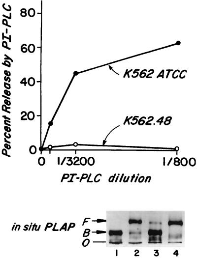
Figure 1.
(A) In situ cleavage by PI-PLC of endogenous DAF in K562 ATCC and K562.48 cells. Cells were treated with PI-PLC and then stained with anti-DAF or nonrelevant mAbs and analyzed by flow cytometry. Percent DAF released equals the difference between the fluorescence of buffer- and PI-PLC-treated cells. (B) ND-PAGE analyses of in situ PLAP protein. Samples in lanes 1 and 2 were treated with buffer at 0°C overnight, and in lanes 3 and 4 treated with hydroxylamine, pH 10.7 at 0°C overnight. Samples in lanes 1 and 3 then were incubated with buffer and samples in lanes 2 and 4 with PI-PLC (1:50) at 37°C for 2 hr. F, protein freed from detergent micelles; B, protein bound to detergent micelles; O, origin.

