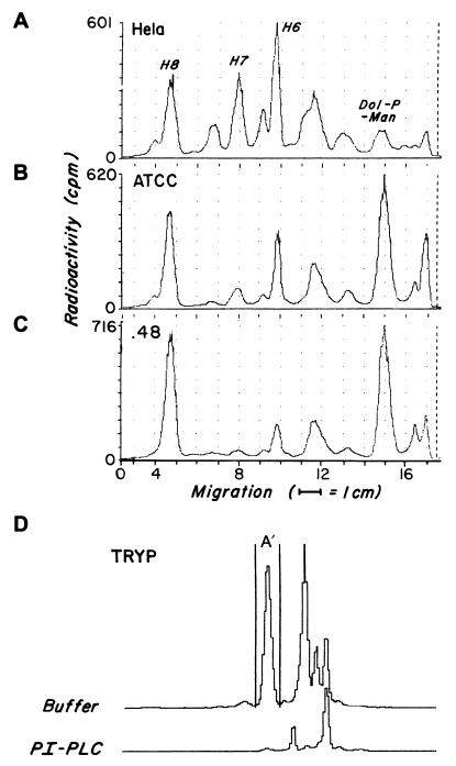
Figure 2.
TLC analyses of GPI products deriving from [3H]mannose labeling of K562 ATCC and .48 lines. [3H]mannose-labeled GPI intermediates from tunicamycin-pretreated HeLa cells (A), K562 ATCC cells (B), or K562.48 cells (C) were separated on TLC plates developed in chloroform:methanol:water (10:10:3). The positions of previously characterized HeLa cell bands H6, H7, and H8 and of dolichol-phosphoryl-mannose are indicated. As control (D) [3H]mannose-labeled Tryp GPIs were prepared from Tryp lysate (1 × 10⁷ cell equivalent) incubated with 10 μCi GDP-[3H]mannose. Aliquots of GPI products deriving from each source alternatively were incubated with buffer or PI-PLC. The radioactivity (cpm) falling within the peak (H8 and H7 for the K562 cell lines and A′ for Tryp) was electronically measured.

