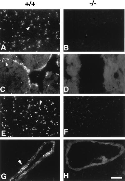
Figure 2.
Immunolocalization of vWf in platelets and tissue sections. Blood smears (A, B, E, and F), heart sections (C and D), or lung sections (G and H) were prepared from wild-type (+/+) and mutant (−/−) mice and stained by immunofluorescence with a polyclonal antibody to mature vWf (A–D) or to the propeptide (E–H). Bright granular staining characteristic of platelet α-granules (A and E), and Weibel–Palade bodies (C and G), stronger than the autofluorescent background of tissue, is observed only in the samples from the wild-type animals (arrowheads). (Bar = 10 μm.)

