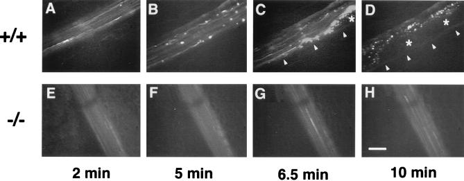
Figure 6.
In vivo thrombosis model in arterioles after ferric chloride-induced injury. An arteriole from a wild-type (+/+) or a vWf-deficient (−/−) mouse was injured by ferric chloride superfusion and photographed at four time points after injury. A progression in the quantity of platelets interacting with the vessel wall is visible in the wild-type arteriole (A–D), leading to complete occlusion and blood stasis in D. Arrowheads in C–D show edge of vessel above which is the forming thrombus. Asterisks indicate the center of a thrombus containing unlabeled and bleached platelets. Almost no platelet interactions are visible in the vWf-deficient arteriole (E–H). (Bar = 50 μm.)

