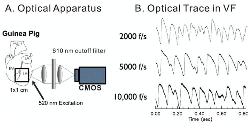
Figure 1.

Optical apparatus. Transmembrane potentials were imaged optically using a 100 × 100 CMOS camera at up to 10,000 frames per second (0.1-ms resolution). A: Orientation of field of view and schematics of optical apparatus. B: Sample traces at 2,000, 5,000, and 10,000 frames per second. Signal-to-noise ratio was 81:1, 47:1, and 21:1 respectively.
