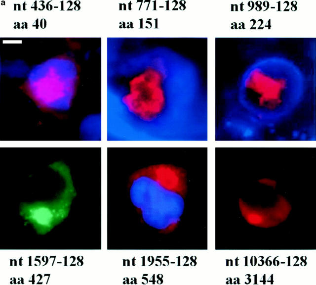
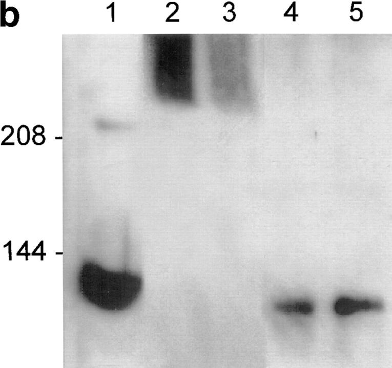
Figure 2.
(a) Immunofluorescence of 293T cells transfected with the 436, 771, 989, 1597, 1955, and 10366 constructs, each containing 128 glutamines. 36–40 h after transfection, the cells were processed for immunofluorescence using mAb 2166 (red) or BKP1 (green) to detect huntingtin. The cells were counterstained with the DNA stain DAPI (blue) to label the nucleus (the DAPI stain is not visible in the cells shown for 1597-128 and 10366-128). Huntingtin located in the nucleus appears pink as the red stain is overlaid with blue. Huntingtin from the 436, 771, and 989 constructs (top row) appears as large intranuclear aggregates. Huntingtin from the 1597, 1955, and 10366 constructs (bottom row) show perinuclear aggregates and diffuse cytoplasmic stain. (b) Size-exclusion chromatography of a lysate from 293T cells transfected with HD1955-128 and treated with tamoxifen. 1-ml fractions were collected at 4°C, and samples were immediately analyzed by reducing SDS-PAGE and Western blotting with BKP1 antibody. Large molecular weight complexes were not retained by the column and flowed through into the void volume. Huntingtin aggregates isolated in the void volume were observed as species that did not denature upon SDS-PAGE and did not exit the stacking gel (V o = 42 ml; lanes 2 and 3). Under the same conditions, no aggregation was observed in the whole cell lysate (lane 1) nor in the monomeric huntingtin fractions (V = 72 ml; lanes 4 and 5). Bar in a: (436-128) 11.3 μm; (771-128) 5.31 μm; (989-128) 11.2 μm; (1597-128) 11.97 μm; (1955-128) 12 μm; (10366-128) 11.4 μm.


