
Figure 4.


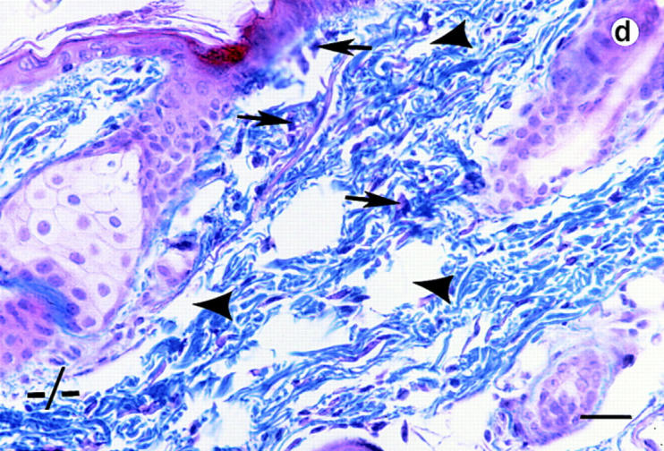
Histological analyses on skin alterations. Sections through skin from dorsal (a and b) and ventral (c and d) surfaces. Wild-type animals (+/+) shown in a and c and lumican null mutants (−/−) shown in b and d. The sections were trichrome stained in which the connective tissue, collagens primarily stain blue. Note disoriented fibroblasts (arrows) and open spaces (arrowheads) in the dermis of b and d. Bar, 50 μm.