
Figure 7.
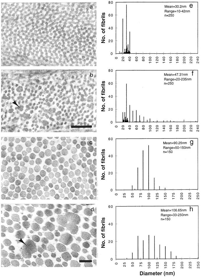
Transmission electron micrography of cornea (a and b) and tail dermis (c and d) collagen preparations (17). (a and c) Homozygous wild type, and (b and d) Lumtm1Sc homozygous. Note larger abnormal shaped fibrils in cross section (arrowhead) in b and d. Fibril diameter frequency (e–h). Homozygous wild-type cornea and tail collagen (e and g) and lumtm1Sc homozygous cornea and tail collagen (f and h). Bar, 0.2 μm.