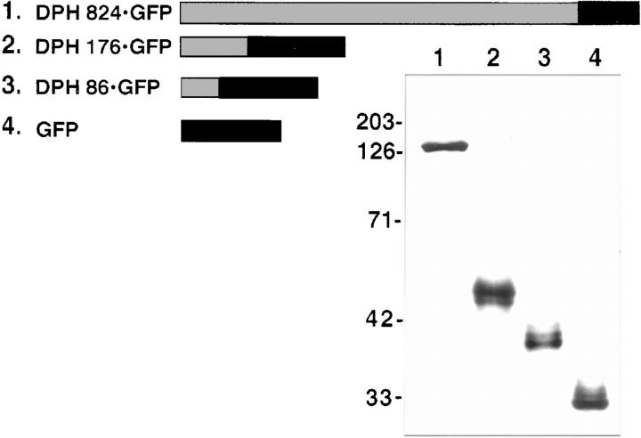
Figure 6.
Construction and expression of DPH fusion proteins. (Top) Diagram indicating fusion proteins generated from portions of DPH (grey) linked to GFP (black bars). (Bottom) Cells were transfected with expression vectors encoding the DPH-GFP fusion proteins. After transfection, extracts were isolated and subjected to electrophoresis through 10% SDS polyacrylamide gels, electroblotted onto nitrocellulose paper, probed with anti-GFP and subjected to chemiluminescence. Molecular mass standards in kD at left.

