
Figure 7.
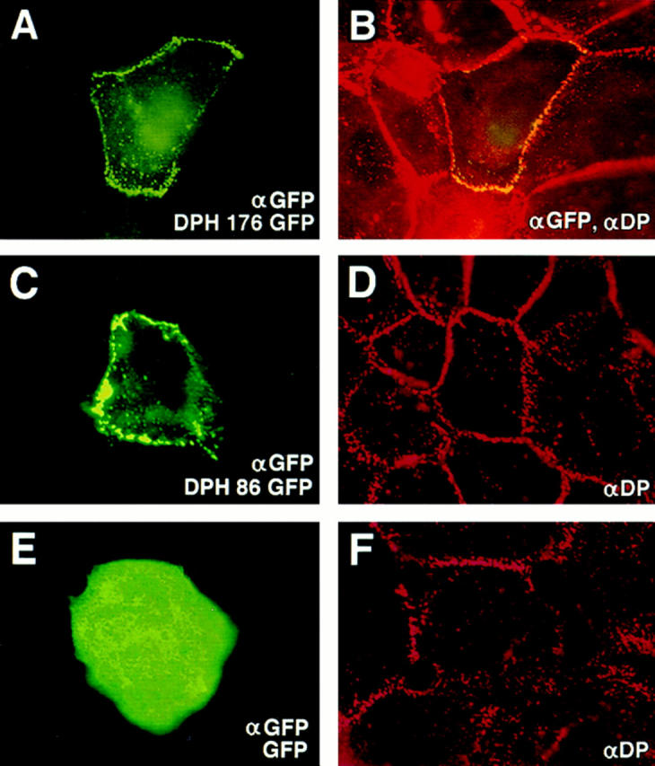
Localization of desmoplakin fusion proteins to desmosomes. Keratinocytes were transfected with expression vectors encoding desmoplakin fusion proteins. After transfection, cells were fixed and stained with the indicated antibodies (lower right). The αDP used was to the tail segment of DP, not present in the fusion protein. Representative cells for each construct were photographed under an 100× objective, and frames are displayed here in pairs, with transgene product staining at left and double-staining at right. Note that cells expressing DPH 176 GFP or DPH 86 GFP displayed predominantly punctate αGFP/αDP staining at the periphery, typical of desmosomal localization; in contrast, cells expressing GFP exhibited cytoplasmic staining with little or no membrane staining.