
Figure 3.
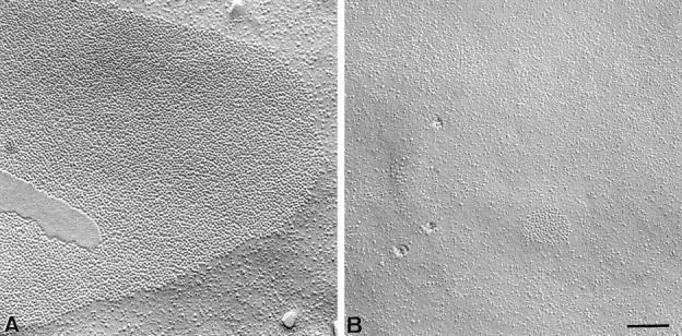

Freeze-fracture analysis of gap junctions in pancreatic acinar cells. In contrast to gap junctions observed in wild-type animals (A), pancreatic acinar cells from Cx32 (−/−) mice showed less frequent gap junctions plaques of smaller areas (B). (C) Quantitative evaluation of gap junctions in wild-type and Cx32 (−/−) pancreatic acinar cells. The number of gap junction plaques was reduced five fold in the pancreas of Cx32 (−/−) mice (open columns) as compared with that evaluated in wild-types (solid columns). The size of gap junction plaques was also reduced twenty fold in Cx32 (−/−) exocrine pancreas. Stars, differences at a P < 0.001 level. Bar, 130 nm.