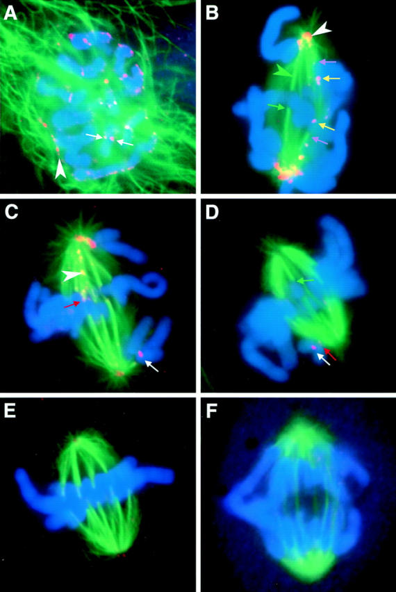
Figure 4.

Mad2 leaves kinetochores as they accumulate kinetochore microtubules. Mitotic PtK1 cells were labeled with antibodies to Mad2 (orange/pink), antibodies to α-tubulin (green), and the DNA stain Hoechst 33342 (blue). (A) Just after nuclear envelope breakdown. White arrows, Mad2 accumulating on kinetochores; white arrowhead, Mad2 on residual nuclear envelope. (B) Early prometaphase. Green arrowhead, kinetochore fibers are visible as bright dense bundles of green microtubules that end abruptly on a chromosome; white arrowhead, spindle pole. (C) Mid-prometaphase. White arrowhead, Mad2 on kinetochore microtubules. (D) Late prometaphase. (E) Metaphase. (F) Anaphase. Yellow arrows, newly attached leading kinetochores on congressing chromosomes label brightly for Mad2; purple arrows, trailing kinetochores on attached kinetochores label less brightly; green arrows, attached kinetochores that lack Mad2; and red arrows, attached kinetochores on which some Mad2 is still visible.
