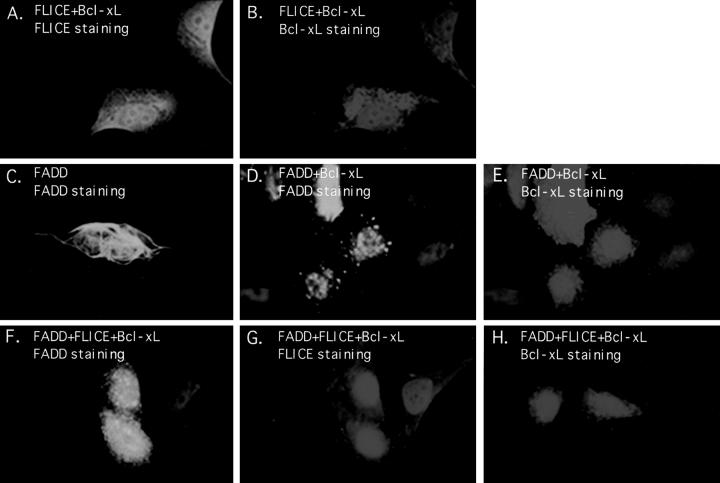
Figure 6.
Bcl-xL attenuates the formation of FADD filaments. HeLa cells were transfected with the expression plasmids indicated and indirect immunofluorescence was performed 48 h posttransfection. HeLa cells were cotransfected with HA-tagged FLICE and Flag-tagged Bcl-xL and then stained with an anti-HA polyclonal antibody (A) or an anti-Flag monoclonal antibody (B). Cells were transfected with AU1-tagged FADD alone and visualized with an anti-FADD polyclonal antibody (C). In D and E, cells were transfected with AU1-tagged FADD and Flag-tagged Bcl-xL and protein expression was detected with an anti-FADD polyclonal antibody (D) and an anti-Flag monoclonal antibody (E). In panels F–H, cells were transfected with AU1-tagged FADD, HA-tagged FLICE, and Flag-tagged Bcl-xL. Proteins were visualized with anti-FADD polyclonal antibody (F), anti-HA monoclonal antibody (G), and anti-Flag monoclonal antibody (H). All cells were also transfected with CrmA to retain cell viability.

